
CNS Resources
The Digestive System of Vertebrates
This information is designed for use by physiologists, zoologists, veterinarians, nutritionists, and others interested in the vertebrate digestive system, and to aid in the teaching of comparative anatomy, physiology, and nutrition.
In 1969, a graduate course in the comparative physiology of the vertebrate digestive system was initiated at the College of Veterinary Medicine of Cornell University. This was followed by a series of graduate courses in comparative anatomy, nutrition, pathology, microbiology, and infectious diseases taught by faculty in the colleges of Veterinary Medicine, Nutrition, and Animal Science. In 1972 a five-year grant from the National Institutes of Health (NIH) was awarded for a Training Program in Comparative Gastroenterology. The co-author of the present website (CES) served as the initial Program Director. The course lectures served as the basis for a text: Comparative Physiology of the Vertebrate Digestive System (Stevens 1988), published by Cambridge University Press. A second edition of this text by Stevens and Hume, with a greater emphasis on interpretation, was published in 1995. The information from this text was, in turn, updated and summarized in a CD-ROM teaching unit entitled The Digestive System of Vertebrates (Stevens 2000).
Esther J. Finegan, PhD - University of Guelph, Ontario, N1G 2W1 Canada
C. Edward Stevens, PhD, DVM - Department of Molecular Biomedical Sciences, College of Veterinary Medicine, North Carolina State University, Raleigh, NC, 27606 USA.
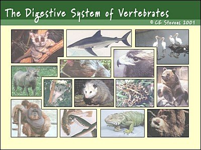

The Digestive System of Vertebrates
Authors:
Esther J. Finegan, PhD - University of Guelph, Ontario, N1G 2W1 Canada.
C. Edward Stevens, PhD, DVM - Department of Molecular Biomedical Sciences, College of Veterinary Medicine, North Carolina State University, Raleigh, NC, 27606 USA.
The vertebrates are comprised of approximately 45,000 species of fish, amphibians, reptiles, birds, and mammals. Their digestive system consists of a headgut, foregut, midgut, hindgut, pancreas, oral glands and billiary system. The headgut (oral cavity and pharynx) serves principally for the procurement and ingestion of food. The foregut consists of the gullet or esophagus and a stomach for the storage and initial digestion of food. The biliary system produces bile salts that aid in lipid digestion by acting as detergents for the emulsification of fat. Food is digested by a compliment of endogenous enzymes secreted by the digestive tract and pancreas tissue or present in the apical membranes and contents of intestinal mucosal cells. The hindgut serves principally for recovery of electrolytes, water, urea-nitrogen and carbohydrates that escape the upper digestive tract. Indigenous bacteria provide an additional source of nutrients.
Glandular and enteric cells secrete mucous and water to aid in digestion of food and absorption of nutrients. Ingesta and digesta are mixed with secretions and moved through the digestive tract by its motor activity. The addition of bile to the digesta increases the digestibility of lipids. Electrolytes, vitamins, and the end products of carbohydrate, and protein digestion are absorbed by transport across intestinal cell membranes. The midgut or small intestine serves as the principal site of digestion and absorption in most vertebrates. Its absorptive surface is increased by microvilli, and may be further expanded by villi, pyloric ceca, spiral valves, ridges, pockets or folds in some species.
Despite its many common characteristics, individual parts of the vertebrate digestive system can demonstrate a wide range of structural and functional variation, both within and among the classes of vertebrates. The headgut and hindgut show a wide range of adaptations to diet and habitat. The stomach is absent in some species of fish and serves as the principle site of microbial fermentation in some mammals. Some fish lack a distinct pancreas and the motor activity of the midgut differs between continuous and discontinuous feeders. The hindgut is relatively short in fish and tends to be longest and most complex in terrestrial herbivores. In addition to these structural differences, species can show wide variation in the degree of enzyme activity and some enzymes are absent from some species.
An understanding of these adaptations is essential for the proper care and maintenance of domesticated and captive animals, and the preservation of wildlife and endangered species. Comparative studies have also provided much of our present understanding of the basic physiological mechanisms and how they evolved. This has led to a constant search for animals that can serve as good models for the study of human physiology and diseases. The contribution of indigenous microorganisms to sodium, nitrogen, water, and energy conservation by the large intestine was first demonstrated in studies of the same phenomena in the ruminant forestomach. Models for the study of disease are generally more obvious, due to similarities in signs, symptoms, or etiology. However, some of the best animal models are the result of their apparent differences rather than similarities. For example, the difference (simplicity) of non glandular frog skin from epithelium provided the basic model for determining the mechanisms of ion transport across the intestinal epithelium.